

Erythroxylum Coca Seeds
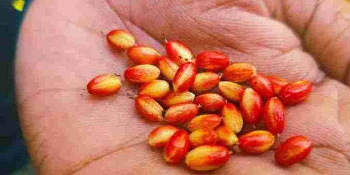
Erythroxylum coca is a little, evergreen bush having a place with the Erythroxylaceae own loved ones. The plant creates little, red berries that consolidate the seeds, which may be oval and level, estimating around three-4 millimetres long. These seeds are encased in a difficult, defensive shell that aids in their dispersal and germination.
The fresh coca seeds are critical for the plant’s replica. Upon maturity, the berries fall to the ground, where they're often eaten up by birds and other animals. The seeds skip through the digestive structures of these animals, which aids their dispersal throughout the woodland ground. This herbal system guarantees the propagation of the species in its native habitat.
Historical Importance of Erythroxylum Coca Seeds
The records of Erythroxylum coca are significantly joined with the lifestyle of South America, essentially the Andean municipal foundations. For heaps of years, native people groups have developed and regarded the coca plant for its restorative, non common, and social utilises. The seeds, despite the fact that they are not habitually noted in memorable records, have played a critical role in safeguarding the progression of those practices.
Archaeological proof suggests that the coca plant became domesticated around 3000 BCE. The seeds have been selectively bred to produce plants with suitable trends, which include higher alkaloid content material and more resistance to pests and illnesses. This early rural practice features the resourcefulness of authentic civic establishments and their significant skill of plant science.
Cultural Significance
One of the most extremely good cultural practices concerning coca seeds is the culture of coca leaf chewing. The leaves are harvested from mature vegetation, which might be grown from carefully decided seeds. This practice, called acullico or mambeo, is deeply ingrained in Andean lifestyle and serves more than one function. It acts as a stimulant, helping individuals deal with the high altitudes of the Andes, and is also used in social and spiritual contexts.
Coca seeds also are vital to the education of conventional drugs. Indigenous healers, known as curanderos, use diverse components of the coca plant, together with the seeds, to treat various illnesses. The seeds are believed to possess recuperation properties which could alleviate digestive troubles, fatigue, and respiration problems. This traditional know-how has been surpassed down thru generations, highlighting the enduring cultural significance of coca seeds.
Cultivation Practices of Erythroxylum Coca Seeds
Developing Erythroxylum coca from seeds is a careful technique that calls for mindful interest. The seeds are generally amassed from ripe berries and dried before planting. This drying technique allows to interrupt the seed dormancy, bearing in mind successful germination.
The planting of coca seeds generally takes place at the onset of the wet season, ensuring that the younger seedlings acquire good enough moisture for boom. The seeds are sown in well-tired soil, often enriched with natural count numbers to offer critical vitamins. Farmers typically plant the seeds in small nurseries earlier than transplanting the younger plants to their final growing locations.
The cultivation of coca flora requires keen information of the neighbourhood surroundings. The vegetation thrive in well-tired, fertile soil and require a warm, humid climate with sufficient rainfall. Farmers ought to additionally protect the young plants from pests and illnesses, that may significantly impact yields. This is regularly done via natural farming practices and traditional information handed down through generations.
Erythroxylum Coca Seeds in the Global Context
While the plant is regularly related to the manufacturing of cocaine, its traditional uses and cultural importance have to not be unnoticed. Efforts to regulate and manipulate the cultivation of coca plant life have had profound impacts at the communities that depend on them.
The global community has taken steps to deal with the complex problems surrounding coca cultivation. Policies aimed at removing coca flowers have often confronted resistance from neighbourhood groups, who argue that these efforts threaten their cultural historical past and financial survival. In response, opportunity development packages were implemented to provide farmers with viable options to coca cultivation.
Despite those challenges, the call for coca products, which include seeds, remains high. The plant’s medicinal houses and cultural significance maintain a power hobby in its cultivation and use. Researchers are also exploring the ability to buy coca seeds in present day remedies, looking to harness their specific residences for healing purposes.
Conclusion
Erythroxylum coca seeds are a critical part of the plant’s life cycle and preserve deep historical, cultural, and economic significance. From their position in ancient Andean civilizations to their location in modern-day society, those seeds constitute a fascinating intersection of botany, history, and tradition. Understanding the significance of coca seeds gives treasured insights into the long-lasting legacy of the coca plant and its region within the world today.